


Breaking News
 SPLC 'Fascism Expert' Funneled $1.2 MILLION in Donor Cash to Her Neo-Nazi Informant/Lover
 Israeli Ministers Say Israel Isn't Bound by US-Iran Deal, Won't Withdraw From Lebanon
 EXCLUSIVE: Top FBI Whistleblower Says The Supposed Terror Plot Targeting The White House...
 Heterofatalism
Top Tech News
 Heads up: Apparently the government is hiding cameras inside fake utility boxes
 Sodium Batteries And EVs That Power The Grid: Inside GM's Big Energy Push
 NUCLEAR ENGINE - UNLIMITED LUXURY - 20 YEARS WITHOUT REFUELING
 China Unveils Nuclear-Powered Floating Hub For Green Shipping
 China Launches World's 1st Commercial Brain Chip, Beating Elon Musk's Neuralink!
 Modular next-gen US nuclear reactor goes critical
 How EMF's cause disease
 This Company Will Add Phone, AirPod, and Smartwatch Trackers to License Plate Readers
 Elon Details SpaceX AI Data Center in Space Details and Roadmap
 5-in-1 miniature surgical robot is the size of a seed


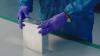
CERN chip enables first 3D color X-ray images of the human body
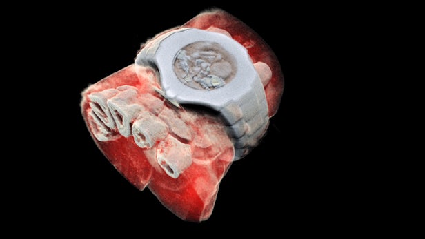
Medical X-ray scans have long been stuck in the black-and-white, silent-movie era. Sure, the contrast helps doctors spot breaks and fractures in bones, but more detail could help pinpoint other problems. Now, a company from New Zealand has developed a bioimaging scanner that can produce full color, three dimensional images of bones, lipids, and soft tissue, thanks to a sensor chip developed at CERN for use in the Large Hadron Collider.
Mars Bioimaging, the company behind the new scanner, describes the leap as similar to that of black-and-white to color photography. In traditional CT scans, X-rays are beamed through tissue and their intensity is measured on the other side. Since denser materials like bone attenuate (weaken the energy) of X-rays more than soft tissue does, their shape becomes clear as a flat, monochrome image.

But for the new technology, which Mars calls "Spectral CT," the sensor can measure the attenuation of specific wavelengths of the X-rays as they pass through different materials. After running the spectroscopic data through specific algorithms, a 3D color image is generated that clearly shows muscle, bone, water, fat, disease markers – and even a watch. The end results are unnerving, like someone's sculpted a detailed clay model of your insides.
At the heart of the Spectral CT scanner is a Medipix3 chip. This device, which detects and counts every individual particle that hits each pixel on the sensor, was originally developed at CERN to precisely track particles in the Large Hadron Collider.
A small version of the device has been tested to see how well it can diagnose bone and joint health, spot cancer, and pick up early markers for vascular diseases. So far, the results have been promising, the team says.
"In all of these studies, promising early results suggest that when spectral imaging is routinely used in clinics it will enable more accurate diagnosis and personalization of treatment," says Anthony Butler, one of the creators of the 3D scanner.