Breaking News
 New Coalition Aims To Ban Vaccine Mandates Across US
 We Are Sleepwalking Into An Apocalyptic War With Iran
 Munich Security Conference and the U.S. Elephant in the Room
 Government's Business Plan Is Predation
Top Tech News
 New Spray-on Powder Instantly Seals Life-Threatening Wounds in Battle or During Disasters
 AI-enhanced stethoscope excels at listening to our hearts
 Flame-treated sunscreen keeps the zinc but cuts the smeary white look
 Display hub adds three more screens powered through single USB port
 We Finally Know How Fast The Tesla Semi Will Charge: Very, Very Fast
 Drone-launching underwater drone hitches a ride on ship and sub hulls
 Humanoid Robots Get "Brains" As Dual-Use Fears Mount
 SpaceX Authorized to Increase High Speed Internet Download Speeds 5X Through 2026
 Space AI is the Key to the Technological Singularity
 Velocitor X-1 eVTOL could be beating the traffic in just a year
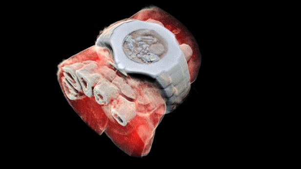
CERN chip enables first 3D color X-ray images of the human body

Sure, the contrast helps doctors spot breaks and fractures in bones, but more detail could help pinpoint other problems. Now, a company from New Zealand has developed a bioimaging scanner that can produce full color, three dimensional images of bones, lipids, and soft tissue, thanks to a sensor chip developed at CERN for use in the Large Hadron Collider.
Mars Bioimaging, the company behind the new scanner, describes the leap as similar to that of black-and-white to color photography. In traditional CT scans, X-rays are beamed through tissue and their intensity is measured on the other side. Since denser materials like bone attenuate (weaken the energy) of X-rays more than soft tissue does, their shape becomes clear as a flat, monochrome image.


