


Breaking News
 THE BUCCANEER (1958) Theatrical Trailer - Yul Brynner, Claire Bloom, Charles Boyer
 Saturday Class - The American Revolution was a Christian Revolution - June 20, 2026
 Why Hasn't There Been a Recession Yet?
 As America Marks Its 250th Anniversary, Debates Rage Over National Identity
Top Tech News
 Our Diesel-Electric Truck Is So Quiet the Military Wants One
 World's first hotel entirely staffed by robots to open in 2027
 Researchers in China are ignoring bug spray, citronella, and netting.
 Our bodies may be able to regrow lost limbs after all
 Chinese cars go blacker than black via hybrid nano tech
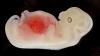 World first: Human embryo model grows its own organs – in the lab
 Dead lithium batteries revived to 95% capacity via electrochemical bath
 Compact laser engraver levels up your DIY crafts setup
 'Groundbreaking' Potential Lupus Cure Sends Patients into Remission, Allowing Dreams...
 SpaceX Orbital Travel and Orbital Hotels Need Starfall – Getting Back Safe and Cheap is Exciting


Magnetic resonance imaging of melanoma exosomes
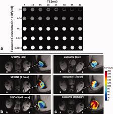
The ability to modify and track exosomes in vivo is essential to understanding exosome pathogenesis, and for utilizing exosomes as effective diagnostic and therapeutic nanocarriers to treat diseases.
Researchers from the Washington University School of Medicine recently reported a new electroporation method that allow exosomes to be loaded with superparamagnetic iron oxide nanoparticles for magnetic resonance tracking. Building on this approach, they now demonstrate for the first time using a C57BL/6 mouse model that melanoma exosomes can be imaged in vitro, and within lymph nodes in vivo with the use of standard MRI approaches.