


Breaking News
 Trump & NATO Kiss and Make Up as Europeans Pledge To Buy More U.S. Weapons
 Special Saturday Broadcast: Trump Just Threatened Iran With Total War If They Try To Assassinate...
 EXCLUSIVE: Los Angeles Gives Free Pass to Illegal Alien Food Vendors While Restaurants Must...
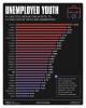 Visualizing Europe's Soaring Youth Unemployment
Top Tech News
 Modular Reactors To Solve Data Center Hysteria?
 DeepSeek Developing In-House AI Chip In Bid To Cut Nvidia Reliance
 America just took three brand-new nuclear reactors critical in thirty days, a first for any...
 Your brain doesn't peak in your 20s after all: Study reveals your mind is at its sharpest betwee
 Compasses, not maps: China is building a different type of AI
 The Return of the Ekranoplan
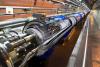 Farewell, atom-smashing Large Hadron Collider
 This is already starting.
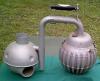 The Crosley IcyBall
 It's Not a Conspiracy Anymore: Med Beds Exist and Trump Knows It


3D color X-ray machine heads for trials
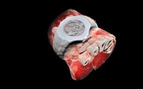
But this new scanner adds color and a third dimension, creating high resolution, cutaway 3D models that can diagnose bone fractures and monitor healing. New Zealand-based Mars Bioimaging (MBI) has now conducted a feasibility study of the machine, with a larger international trial set to begin soon.
In a traditional CT scan, X-rays are beamed through the target area of the body, and the radiation is absorbed more readily by denser tissues like bone, while passing more easily through softer tissues. The end result is that high contrast black-and-white image we know so well.
But the new technology collects more nuanced data about how the X-rays are absorbed by different tissues. It's built around a chip called the Medipix3, which tracks every photon that hits every pixel on the sensor, and processes their interactions with various atoms in the body. By doing so, it can determine the density and composition of those tissues more accurately.