


Breaking News
 Americans Are Officially Out Of Money To Spend -- We Had To React
 Arizona reports several cyclosporiasis cases: What to know | FOX 10 Phoenix
 South Korea's AI Bubble Just Popped
 It's Not Just a New Jersey Scandal – SIX States Were Previously Caught Including THOUSANDS...
Top Tech News
 Anthropic is launching its own drug discovery programs for rare diseases using Claude...
 SpaceX AI Satellites Will Have 250 Kilowatts of Power
 Chinese researchers have developed a sodium-metal battery that can fully charge in just 4 minutes...
 SpaceX Starship Flight 13 in 3 Days - Thursday July 13
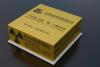 Chinese Scientists Develop Nuclear Battery Using Carbon-14
 Teleoperated humanoid robots complete first-ever live surgery
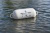 Floating capsule auto-disinfects water without chemicals or battery
 Modular Reactors To Solve Data Center Hysteria?
 DeepSeek Developing In-House AI Chip In Bid To Cut Nvidia Reliance
 America just took three brand-new nuclear reactors critical in thirty days, a first for any...


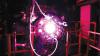
Live view of mysterious Parkinson's protein points to new treatments

The team has produced an unprecedented "live action" view that shows how this protein is activated, providing researchers with a blueprint for therapies that help prevent cell death associated with the condition.
Parkinson's disease takes hold when nerve cells in the brain responsible for producing the chemical dopamine die off or become impaired, but the mechanisms behind this process has remained unclear. Research has pointed to the role mitochondria might play, as the power plants that provide cells with chemical energy, and how that role might take on a sinister nature when mitochondria begin to malfunction.
As we age, mitochondria become damaged and build up in the body, with studies showing how they can change shape and themselves create a toxic environment for diseases like Parkinson's and Alzheimer's to take hold. Research has shown that a protein called PINK1 plays an important protective role in the face of this threat, by tagging damaged mitochondria for destruction and removal, enabling them to be replaced with healthy mitochondria instead.
But when there are defects in the PINK1 protein or other components of this important pathway, the mitochondria are unable to be recycled and cells become starved of energy. In this way, PINK1 has been known to play an important role in the early onset of Parkinson's disease, and scientists have been able to capture differing images of they way it is activated. Now scientists at the Walter and Eliza Hall Institute of Medical Research have used cutting-edge cryo-electron microscopy technology to observe the protein in "exquisite molecular detail" and join together the different pieces of the puzzle.