


Breaking News
 'KICKBACK' SCHEME: Nick Shirley EXPOSES alleged NYC senior daycare center fraud
 Oil prices are in free fall and could keep going to $40. Which implies $2 gasoline.
 Can 'Spaceballs 2' Be As Good As Its Marketing Campaign?
 He Risked Everything To Warn You: No One Is Ready For What's Coming...
Top Tech News
 Modular Reactors To Solve Data Center Hysteria?
 DeepSeek Developing In-House AI Chip In Bid To Cut Nvidia Reliance
 America just took three brand-new nuclear reactors critical in thirty days, a first for any...
 Your brain doesn't peak in your 20s after all: Study reveals your mind is at its sharpest betwee
 Compasses, not maps: China is building a different type of AI
 The Return of the Ekranoplan
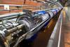 Farewell, atom-smashing Large Hadron Collider
 This is already starting.
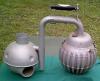 The Crosley IcyBall
 It's Not a Conspiracy Anymore: Med Beds Exist and Trump Knows It


'Theranostics' approach seeks and destroys deadly pancreatic cancer

The one-two punch provided by the novel approach could pave the way for earlier detection and more effective treatment of the disease.
With an average five-year survival rate of less than 10%, pancreatic ductal adenocarcinoma (PDAC) is one of the most lethal forms of cancer. It's also difficult to detect using conventional imaging methods, including positron emission tomography (PET) scans.
Now, researchers at Osaka University in Japan have developed a strategy for combatting this deadly cancer by combining therapeutics and diagnostics – 'theranostics' – into a single, integrated process.
The process developed by the researchers uses radioactive monoclonal antibodies (mAb) to target glypican-1 (GPC1), a protein highly expressed in PDAC tumors. GPC1 has been implicated in cancer cell proliferation, invasion, and metastasis, and high expression of the protein is a poor prognostic factor in some cancers, including pancreatic cancer.
"We decided to target GPC1 because it is overexpressed in PDAC but is only present in low levels in normal tissues," said Tadashi Watabe, the study's lead author.
The researchers injected human pancreatic cancer cells into mice, allowing them to develop into a full tumor. The xenograft mice were administered intravenous GPC1 mAb labeled with radioactive zirconium (89Zr) and observed for antitumor effects.
"We monitored 89Zr-GPC1 mAb internalization over seven days with PET scanning," said Kazuya Kabayama, the study's second author. "There was strong uptake of the mAb into the tumors, suggesting that this method could support tumor visualization. We confirmed that this was mediated by its binding to GPC1, as the xenograft model that had GPC1 expression knocked out showed significantly less uptake."