


Breaking News
 SPLC 'Fascism Expert' Funneled $1.2 MILLION in Donor Cash to Her Neo-Nazi Informant/Lover
 Israeli Ministers Say Israel Isn't Bound by US-Iran Deal, Won't Withdraw From Lebanon
 EXCLUSIVE: Top FBI Whistleblower Says The Supposed Terror Plot Targeting The White House...
 Heterofatalism
Top Tech News
 Heads up: Apparently the government is hiding cameras inside fake utility boxes
 Sodium Batteries And EVs That Power The Grid: Inside GM's Big Energy Push
 NUCLEAR ENGINE - UNLIMITED LUXURY - 20 YEARS WITHOUT REFUELING
 China Unveils Nuclear-Powered Floating Hub For Green Shipping
 China Launches World's 1st Commercial Brain Chip, Beating Elon Musk's Neuralink!
 Modular next-gen US nuclear reactor goes critical
 How EMF's cause disease
 This Company Will Add Phone, AirPod, and Smartwatch Trackers to License Plate Readers
 Elon Details SpaceX AI Data Center in Space Details and Roadmap
 5-in-1 miniature surgical robot is the size of a seed


MIT imaging technique sheds light on the brain's electrical activity
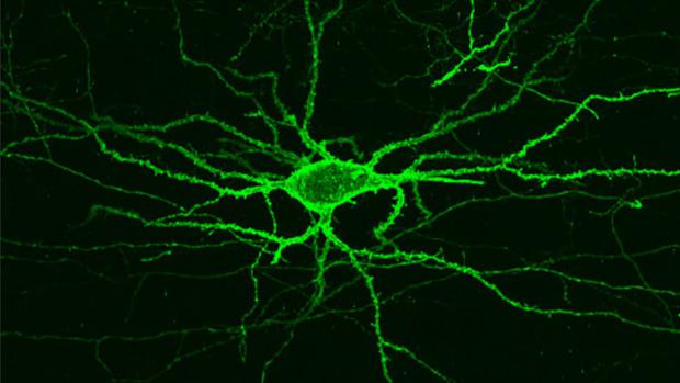
Brain MRIs offer important insight into how our brains work, but they can only produce crude approximations of the areas that are activated by a given stimulus. In order to unravel the minutiae of how neurons communicate and collaborate to form thoughts and feelings, we would need imaging tools with vastly improved resolutions.
Today, far from being able to tackle the 86 billion neurons in the human brain, neuroscientists must settle for studying simple organisms like worms and fish larvae (with neuron counts in the hundreds), relying on slow and cumbersome methods like implanting electrodes into brain tissue to detect electrical signals.
This, however, could soon change. The group of researchers led by Prof. Ed Boyden at MIT has built on previous work to perfect an imaging technique that provides a much fuller picture of the brain's activity. When exposed to red light, a carefully selected fluorescent protein bound to the cellular membrane of neurons reacts to electrical signals by lighting up, to reveal the exact neural path of a thought.