


Breaking News
 IRS Threatens to Seize Tennessee Man's Farm Over Interest Charged on Accidental Refund...
 STIFFED BY THE OBAMAS: Plumbing Subcontractor Forced to Shut Down and Lay Off 25 Union...
 DIGITAL ID FOR TRADES: Mark Carney's SURVEILLANCE NET Closes In!!
 ARE THEY INSANE? Now War With CHINA and RUSSIA??
Top Tech News
 Anthropic is launching its own drug discovery programs for rare diseases using Claude...
 SpaceX AI Satellites Will Have 250 Kilowatts of Power
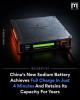 Chinese researchers have developed a sodium-metal battery that can fully charge in just 4 minutes...
 SpaceX Starship Flight 13 in 3 Days - Thursday July 13
 Chinese Scientists Develop Nuclear Battery Using Carbon-14
 Teleoperated humanoid robots complete first-ever live surgery
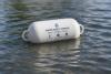 Floating capsule auto-disinfects water without chemicals or battery
 Modular Reactors To Solve Data Center Hysteria?
 DeepSeek Developing In-House AI Chip In Bid To Cut Nvidia Reliance
 America just took three brand-new nuclear reactors critical in thirty days, a first for any...


Portable X-ray device would let patients check their own bones
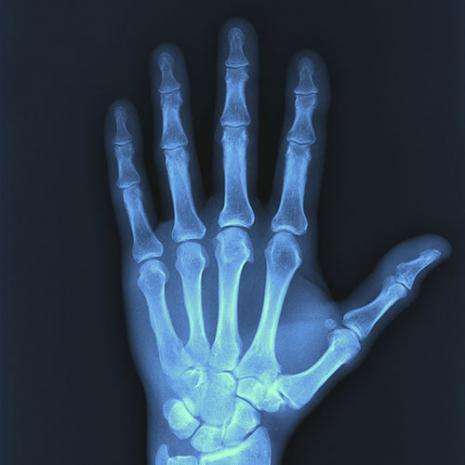
Currently under development at Finland's University of Oulu, the prototype portable X-ray machine measures just 50 by 50 by 130 cm (19.7 by 19.7 by 51.2 in).
Not only is it much smaller than conventional X-ray systems, but because it incorporates built-in radiation shielding, it doesn't have to kept in a lead-lined room, nor does it have to be operated from a separate area. In fact, it utilizes a video screen to guide patients through the process, showing them how and where to place the injured appendage. It then automatically takes the X-rays, and tells the user if a break is detected.
Its instructions – and its imaging voltage – are currently set up for X-raying bones in the palm and ankle. More regions will be added as the system is developed further.
The idea behind the technology is that the relatively inexpensive machines could be set up at locations such as ski resorts or medical clinics, where patients could self-check their injuries to see if a bone was indeed broken. This would reduce the demands placed on larger, pricier, more sophisticated X-ray systems (and their operators), increasing their availability for more important tasks.