


Breaking News
 Americans Are Officially Out Of Money To Spend -- We Had To React
 Arizona reports several cyclosporiasis cases: What to know | FOX 10 Phoenix
 South Korea's AI Bubble Just Popped
 It's Not Just a New Jersey Scandal – SIX States Were Previously Caught Including THOUSANDS...
Top Tech News
 Anthropic is launching its own drug discovery programs for rare diseases using Claude...
 SpaceX AI Satellites Will Have 250 Kilowatts of Power
 Chinese researchers have developed a sodium-metal battery that can fully charge in just 4 minutes...
 SpaceX Starship Flight 13 in 3 Days - Thursday July 13
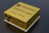 Chinese Scientists Develop Nuclear Battery Using Carbon-14
 Teleoperated humanoid robots complete first-ever live surgery
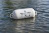 Floating capsule auto-disinfects water without chemicals or battery
 Modular Reactors To Solve Data Center Hysteria?
 DeepSeek Developing In-House AI Chip In Bid To Cut Nvidia Reliance
 America just took three brand-new nuclear reactors critical in thirty days, a first for any...


The Immense Complexity of a Brain is Mapped in 3D for the First Time:
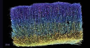
During the last seven years, a global team of more than 150 scientists collaborated on the most complicated neuroscience experiment ever attempted—and they've released their findings this week.
From a tiny sample of tissue no larger than a grain of sand, the MICrONS Project completed the first step toward the goal once thought unattainable: building a functional wiring diagram of a portion of the brain.
Now, they've published their findings in Nature with a collection of ten studies. The 3D wiring diagram and its data are massive—1.6 petabytes in size (equivalent to 22 years of non-stop HD video). They offer a never-before-seen insight into brain function and organization of the visual system.
The research started at Baylor College of Medicine where scientists used specialized microscopes to record the brain activity from a one cubic millimeter portion of a mouse's visual cortex while the animal watched various movies and YouTube clips.
Afterwards, Allen Institute researchers took that same cubic millimeter of the brain and shaved it into more than 25,000 layers, each 1/400th the width of a human hair, and used an array of electron microscopes to take high-resolution pictures of each slice.
By the end, the MICrONS Project—Machine Intelligence from Cortical Networks—built the most detailed wiring diagram of a mammalian brain to date—and it's freely available online.
"A watershed moment for neuroscience, comparable to the Human Genome Project" is the description from David Markowitz, Ph.D., who coordinated this work after leaving the IARPA, the US Intelligence Advanced Research Projects Activity, which partially funded it.
Another team at Princeton University used artificial intelligence and machine learning to reconstruct the cells and connections into a 3D volume. Combined with the recordings of brain activity, it contains 523 million synapses (the connection points between 200,000 cells) and a length of four kilometers of axons (the branches that reach out to other cells).
"Inside that tiny speck is an entire architecture like an exquisite forest," said Clay Reid, Ph.D., senior investigator and one of the early founders of electron microscopy connectomics who brought this area of science to the Allen Institute 13 years ago.
"It has all sorts of rules of connections that we knew from various parts of neuroscience—and within the reconstruction itself, we can test the old theories and hope to find new things that no one has ever seen before."