


Breaking News
 Starship's Thirteenth Flight Test
 Episode 485: THE PEPTIDE DIVIDE: HYPE, HOPE & HARD TRUTHS
 With Lena Petrova
 Second Circuit Upholds New York Vaccine Mandate Against the Amish
Top Tech News
 Anthropic is launching its own drug discovery programs for rare diseases using Claude...
 SpaceX AI Satellites Will Have 250 Kilowatts of Power
 Chinese researchers have developed a sodium-metal battery that can fully charge in just 4 minutes...
 SpaceX Starship Flight 13 in 3 Days - Thursday July 13
 Chinese Scientists Develop Nuclear Battery Using Carbon-14
 Teleoperated humanoid robots complete first-ever live surgery
 Floating capsule auto-disinfects water without chemicals or battery
 Modular Reactors To Solve Data Center Hysteria?
 DeepSeek Developing In-House AI Chip In Bid To Cut Nvidia Reliance
 America just took three brand-new nuclear reactors critical in thirty days, a first for any...


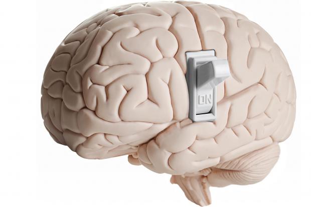
'Sugar switch' in the brain offers new path to treating depression
Scientists from South Korea's Institute for Basic Science (IBS) found that prolonged stress changes how proteins in the medial prefrontal cortex (mPFC) are "decorated" with sialic acid – a sugar molecule that helps shape the surface properties of neurons. These sugar chains, called glycans, are attached after proteins are made, forming the process known as glycosylation. Glycosylation has been studied in terms of how it impacts cancer development and, more recently, neurodegeneration.
One type of glycosylation is O-glycosylation, where sugars attach to oxygen atoms on certain amino acids in a protein. This molecular "sugar coating" helps regulate how neurons connect and signal to one another. Until recently, it was largely overlooked in mental-health research, but scientists are now finding that stress can disturb these sugar patterns, potentially rewiring "normal" communication between brain cells.
In this study, the researchers identified that a single enzyme, St3gal1, performs the final "sugar-capping" step in the process, and this small but integral stage influences how long proteins last and how they interact at synapses – and how, if this falters, it appears to influence depression-like behaviors as a result.
Researchers used high-performance mass spectrometry to map O-glycosylation patterns across nine brain regions in healthy mice. Each area had a distinct sugar signature, reflecting its unique cellular blueprint. When the team compared these with those from chronically stressed mice, they found a significant difference in the prefrontal cortex, a region linked to mood regulation. Here, stress led to a noticeable reduction in that final O-glycosylation sugar-capping step, and a corresponding drop in St3gal1 expression.
Knocking out St3gal1 in healthy mice caused depressive symptoms, including loss of motivation and heightened anxiety. Increasing St3gal1 in stressed mice had the opposite effect, easing those behaviors. This showed that the enzyme has a key role in how stress triggers depression-like changes in the brain. The team also found that St3gal1 helps maintain sugar tags on neurexin-2, a protein that supports communication between neurons. In stressed mice, those tags vanished – along with normal neural signaling – but restoring St3gal1 brought them back.
"This study demonstrates that abnormal glycosylation in the brain is directly connected to the onset of depression," said research fellow Boyoung Lee. "It provides an important foothold for identifying new diagnostic markers and therapeutic targets beyond neurotransmitters."
While the findings were only demonstrated in the brains of male mice – and, of course, the neural networks in humans are far more complex – it provides a new angle for research into depression and its treatment. Many current antidepressants act on serotonin – boosting its levels or altering its signaling – but there's growing evidence suggesting that it's not simply a case of "too little serotonin."